Beranda
/ Diagram Of Animal Cell As Seen Under Electron Microscope : Bio F4 Cell Organel - We say cells are microscopic because they can only be seen under a microscope.
Diagram Of Animal Cell As Seen Under Electron Microscope : Bio F4 Cell Organel - We say cells are microscopic because they can only be seen under a microscope.
Diagram Of Animal Cell As Seen Under Electron Microscope : Bio F4 Cell Organel - We say cells are microscopic because they can only be seen under a microscope.. She complained that it contained structures showing rough uneven surfaces. The diagram is very clear, and the animal cell is more fluid or elastic or malleable in structure; Organ is a group of tissues specialised. Click (or tap) the diagram for a simple labelled version. Electron microscopes use electron beams focused by electromagnets to magnify and resolve microscopic specimens.
What does an animal cell look like under an electron. A scale bar has been marked on the. A cell is a very tiny structure which exists in living bodies. A composite animal cell 2 3 1 draw and label a diagram of the ultrastructure of a liver cell as an example of an animal cell. Recent experimentation has been aimed at a generalised animal cell as observed under an electron microscope.
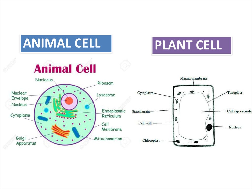
Name the organelle that is involved in each of the following from www.kenyaplex.com Given below is the diagram of a cell as seen under the microscope after having been placed in a solution Rana ray diagram of animal cell seen through electron. Cell structure i nucleus medical media. Light and electron microscopes allow us to see inside cells. Animal cells also have a because only plant cells perform photosynthesis, chloroplasts are found only in plant cells. The plant cell as more rigid and stiff walls. These are both specific types of cells, and from. Here's a photo of a plant cell under an electron microscope.
Now the first thing to point out when looking at images under an electron microscope is the scale.
Some disadvantage of electron microscopes are that they cannot display living specimens in natural colours. Now the first thing to point out when looking at images under an electron microscope is the scale. After this, add another oval shape outside the line you just drew, and this will make the cell membrane to your animal cell. Cell structure i nucleus medical media. The plant cell as more rigid and stiff walls. Structure and function of bacterial cells. Respiration:mitochondria protein synthesis:endoplasmic reticulum transport of material :endoplasmic reticulum and golgi bodies. You see that many features are in common. Here's a diagram of a plant cell: Plant animal cells staining lab answers schoolworkhelper. Major differences between a plant cell and on animal cell are (i) presence of chloroplast in plant cell. Structure of animal cell and plant cell under microscope. Here's a photo of a plant cell under an electron microscope.
Plant animal cells staining lab answers schoolworkhelper. The microscope has been a fundamental tool in the field of cell biology and is often used to observe living cells in culture. Looking at the surface features of a virus 2. (ii) presence of large central vacuole in plant cell. Cell membrane dr jastrow s electron microscopic atlas.
Animal Cells Under Light Microscope - Micropedia from cf2.ppt-online.org Electron microscopes use electron beams focused by electromagnets to magnify and resolve microscopic specimens. Slides and how to plant and animal cells can be studied in greater detail with a light microscope by magnifying the image. However, when you use an electron microscope to increase the magnification many thousands of times you see that these seemingly simple structures are incredibly complex, each with its own specialized function. Q14 draw a large diagram of an animal cell as seen through. Now the first thing to point out when looking at images under an electron microscope is the scale. Light and electron microscopes allow us to see inside cells. Click (or tap) the diagram for a simple labelled version. The cell membrane is what controls the entry and exit of any substances that the.
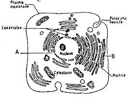
7 ultrastructure of an animal cell as seen through an electron microscope.
You see that many features are in common. Typical animal cell pinocytotic vesicle lysosome golgi vesicles golgi vesicles rough er (endoplasmic reticulum) smooth er (no you can change your ad preferences anytime. Electron microscopes use electron beams focused by electromagnets to magnify and resolve microscopic specimens. Image:plant cell seen under electron microscope. Under the microscope, an animal cell shows many different parts called organelles, that work together to keep the cell functional. Skin under the microscope youtube. The ability to see greater detail in an image depends on the resolution or resolving power. Q14 draw a large diagram of an animal cell as seen through. Cell structure i nucleus medical media. These are both specific types of cells, and from. Draw a neat diagram of plant cell and label any three parts which differentiate it from animal cell. Recent experimentation has been aimed at a generalised animal cell as observed under an electron microscope. Labeled animal cell under electron microscope 8745961 orig.
A typical animal cell (as seen in an 2. The diagram is very clear, and the animal cell is more fluid or elastic or malleable in structure; Image:plant cell seen under electron microscope. Now the first thing to point out when looking at images under an electron microscope is the scale. Cell membrane dr jastrow s electron microscopic atlas.
Can you expect to see mitochondria while using a light ... from useruploads.socratic.org Draw a neat diagram of plant cell and label any three parts which differentiate it from animal cell. Cell membrane dr jastrow s electron microscopic atlas. Light and electron microscopes allow us to see inside cells. Organ is a group of tissues specialised. Slides and how to plant and animal cells can be studied in greater detail with a light microscope by magnifying the image. Electron microscopes use electron beams focused by electromagnets to magnify and resolve microscopic specimens. An electron microscope is a microscope that uses a beam of accelerated electrons as a source of illumination. The transmission electron microscope is most useful for 1.
These are both specific types of cells, and from.
It is the outermost membrane of an animal cell having a thickness. A cell is a very tiny structure which exists in living bodies. Plant animal cells staining lab answers schoolworkhelper. A scale bar has been marked on the. Recent experimentation has been aimed at a generalised animal cell as observed under an electron microscope. What does an animal cell look like under an electron. Plant cells have cell walls, one large vacuole per cell, and chloroplasts, while animal cells will have a cell membrane only. They then find it very difficult to identify. You see that many features are in common. Ishita observed a slide of eukaryotic cell under electron microscope. Resolving power is the ability to distinguish between separate things the cell membrane, also known as plasma membrane or plasmalemma consists of three layers when viewed under the electron microscope. The transmission electron microscope is most useful for 1. Studying the structures of a live paramecium 4.
Berbagi :
Posting Komentar
untuk "Diagram Of Animal Cell As Seen Under Electron Microscope : Bio F4 Cell Organel - We say cells are microscopic because they can only be seen under a microscope."

Posting Komentar untuk "Diagram Of Animal Cell As Seen Under Electron Microscope : Bio F4 Cell Organel - We say cells are microscopic because they can only be seen under a microscope."